Unraveling The Secrets: Uncovering The Forces Behind Chromatid Movement And Anchoring
During cell division, structures called microtubules "move" structures in the cell called chromatids. The spindle fibers, which are composed of microtubules, anchor to the kinetochore of each chromatid and pull them apart to opposite ends of the cell during anaphase.
This process ensures that each new cell receives a complete set of chromosomes and is essential for the proper development and functioning of organisms.
Additional information:
- Kinetochore is a protein complex that assembles at the centromere of each chromosome and serves as the attachment point for spindle fibers.
- Microtubules are long, thin, hollow structures made of tubulin protein. They are part of the cell's cytoskeleton and play essential roles in cell division, cell shape, and intracellular transport.
- Spindle fibers are bundles of microtubules that form during cell division to separate the chromosomes.
What Moves the Chromatids During Cell Division? What Organelle Anchors These?
During cell division, the replicated chromosomes, known as chromatids, must be separated and distributed equally to the daughter cells. Microtubules are responsible for this movement, and the kinetochore is the organelle that anchors the microtubules to the chromatids.
- Kinetochore: protein complex that assembles at the centromere of each chromosome
- Microtubule: long, thin, hollow structures made of tubulin protein
- Spindle fibers: bundles of microtubules that form during cell division to separate the chromosomes
- Motor proteins: move along the microtubules, pulling the chromosomes apart
- Centromere: the region of the chromosome where the kinetochore assembles
- Kinetochore fibers: the microtubules that attach to the kinetochore
- Non-kinetochore fibers: the microtubules that do not attach to the kinetochore
- Astral microtubules: the microtubules that extend from the centrosomes and help to organize the spindle fibers
The interaction of these components is essential for the accurate segregation of chromosomes during cell division. Errors in this process can lead to aneuploidy, which is a condition in which cells have an abnormal number of chromosomes. Aneuploidy can cause a variety of developmental problems and diseases, including cancer.
Kinetochore
The kinetochore is a protein complex that assembles at the centromere of each chromosome. It is the structure to which the spindle fibers attach during cell division. The kinetochore is essential for the accurate segregation of chromosomes during cell division. Errors in kinetochore function can lead to aneuploidy, which is a condition in which cells have an abnormal number of chromosomes. Aneuploidy can cause a variety of developmental problems and diseases, including cancer.
The kinetochore is a complex structure that is composed of over 100 different proteins. These proteins work together to attach the spindle fibers to the chromosome and to ensure that the chromosomes are properly segregated during cell division.
The kinetochore is a vital component of the cell division machinery. It is essential for the accurate segregation of chromosomes during cell division. Errors in kinetochore function can lead to aneuploidy, which is a condition in which cells have an abnormal number of chromosomes. Aneuploidy can cause a variety of developmental problems and diseases, including cancer.
Microtubule
Microtubules are long, thin, hollow structures made of tubulin protein. They are part of the cell's cytoskeleton and play essential roles in cell division, cell shape, and intracellular transport. In the context of cell division, microtubules are responsible for moving the chromatids to opposite ends of the cell.
Each chromatid is attached to a spindle fiber, which is a bundle of microtubules. The spindle fibers shorten, pulling the chromatids apart. This process ensures that each new cell receives a complete set of chromosomes.
Microtubules are essential for the accurate segregation of chromosomes during cell division. Errors in microtubule function can lead to aneuploidy, which is a condition in which cells have an abnormal number of chromosomes. Aneuploidy can cause a variety of developmental problems and diseases, including cancer.
Spindle fibers
Spindle fibers play a central role in moving the chromatids during cell division. They are bundles of microtubules that form during cell division and attach to the kinetochore of each chromatid. The kinetochore is a protein complex that assembles at the centromere of each chromosome.
- Components of spindle fibers
Spindle fibers are composed of microtubules, which are long, thin, hollow structures made of tubulin protein. Microtubules are part of the cell's cytoskeleton and play essential roles in cell division, cell shape, and intracellular transport.
- Formation of spindle fibers
Spindle fibers form during cell division when the centrosomes, which are the microtubule-organizing centers of the cell, move to opposite poles of the cell. The microtubules then polymerize and form bundles that extend from one centrosome to the other.
- Attachment to kinetochores
Spindle fibers attach to the kinetochores of the chromosomes. The kinetochore is a protein complex that assembles at the centromere of each chromosome. The kinetochore serves as the attachment point for the spindle fibers and ensures that the chromosomes are properly segregated during cell division.
- Role in chromosome segregation
Spindle fibers play a critical role in chromosome segregation during cell division. The spindle fibers shorten, pulling the chromatids apart and moving them to opposite poles of the cell. This process ensures that each new cell receives a complete set of chromosomes.
Spindle fibers are essential for the accurate segregation of chromosomes during cell division. Errors in spindle fiber function can lead to aneuploidy, which is a condition in which cells have an abnormal number of chromosomes. Aneuploidy can cause a variety of developmental problems and diseases, including cancer.
Motor proteins
Motor proteins are essential for moving the chromatids during cell division. They attach to the microtubules of the spindle fibers and use energy from ATP to move along the microtubules, pulling the chromatids apart. This process is essential for ensuring that each new cell receives a complete set of chromosomes.
Without motor proteins, the chromosomes would not be able to move apart during cell division. This would lead to aneuploidy, which is a condition in which cells have an abnormal number of chromosomes. Aneuploidy can cause a variety of developmental problems and diseases, including cancer.
Motor proteins are a vital part of the cell division machinery. They play an essential role in ensuring that each new cell receives a complete set of chromosomes.
Centromere
The centromere is the region of the chromosome where the kinetochore assembles. The kinetochore is a protein complex that serves as the attachment point for the spindle fibers during cell division. Spindle fibers are composed of microtubules, which are long, thin, hollow structures made of tubulin protein.
- Role of the centromere in chromosome segregation
The centromere is essential for the accurate segregation of chromosomes during cell division. The kinetochore, which assembles at the centromere, is the attachment point for the spindle fibers. The spindle fibers then shorten, pulling the chromatids apart and moving them to opposite poles of the cell. This process ensures that each new cell receives a complete set of chromosomes.
- Errors in centromere function
Errors in centromere function can lead to aneuploidy, which is a condition in which cells have an abnormal number of chromosomes. Aneuploidy can cause a variety of developmental problems and diseases, including cancer.
The centromere is a vital part of the cell division machinery. It is essential for the accurate segregation of chromosomes during cell division. Errors in centromere function can lead to aneuploidy, which is a condition in which cells have an abnormal number of chromosomes. Aneuploidy can cause a variety of developmental problems and diseases, including cancer.
Kinetochore fibers
Kinetochore fibers are essential for moving the chromatids during cell division. They are microtubules that attach to the kinetochore, which is a protein complex that assembles at the centromere of each chromosome. The kinetochore fibers then shorten, pulling the chromatids apart and moving them to opposite poles of the cell. This process ensures that each new cell receives a complete set of chromosomes.
- Role of kinetochore fibers in chromosome segregation
Kinetochore fibers play a critical role in chromosome segregation during cell division. They are responsible for pulling the chromatids apart and moving them to opposite poles of the cell. This process ensures that each new cell receives a complete set of chromosomes.
- Errors in kinetochore fiber function
Errors in kinetochore fiber function can lead to aneuploidy, which is a condition in which cells have an abnormal number of chromosomes. Aneuploidy can cause a variety of developmental problems and diseases, including cancer.
- Kinetochore fibers and the cell cycle
Kinetochore fibers are essential for the proper progression of the cell cycle. They are involved in the formation of the mitotic spindle, which is a structure that helps to segregate the chromosomes during cell division.
Kinetochore fibers are essential for the accurate segregation of chromosomes during cell division. Errors in kinetochore fiber function can lead to aneuploidy, which is a condition in which cells have an abnormal number of chromosomes. Aneuploidy can cause a variety of developmental problems and diseases, including cancer.
Non-kinetochore fibers
Non-kinetochore fibers are essential for moving the chromatids during cell division. They are microtubules that do not attach to the kinetochore, but they play a vital role in the formation and function of the mitotic spindle. The mitotic spindle is a structure that helps to segregate the chromosomes during cell division.
Non-kinetochore fibers interact with the kinetochore fibers to form the mitotic spindle. The kinetochore fibers attach to the kinetochore, which is a protein complex that assembles at the centromere of each chromosome. The non-kinetochore fibers attach to the poles of the spindle. When the spindle fibers shorten, they pull the chromatids apart and move them to opposite poles of the cell. This process ensures that each new cell receives a complete set of chromosomes.
Non-kinetochore fibers are essential for the accurate segregation of chromosomes during cell division. Errors in non-kinetochore fiber function can lead to aneuploidy, which is a condition in which cells have an abnormal number of chromosomes. Aneuploidy can cause a variety of developmental problems and diseases, including cancer.
Astral microtubules
Astral microtubules are essential for moving the chromatids during cell division. They are microtubules that extend from the centrosomes, which are the microtubule-organizing centers of the cell, and help to organize the spindle fibers. The spindle fibers are bundles of microtubules that attach to the kinetochores of the chromosomes. The kinetochore is a protein complex that assembles at the centromere of each chromosome. When the spindle fibers shorten, they pull the chromatids apart and move them to opposite poles of the cell. This process ensures that each new cell receives a complete set of chromosomes.
Astral microtubules play a vital role in the formation and function of the mitotic spindle. The mitotic spindle is a structure that helps to segregate the chromosomes during cell division. Errors in astral microtubule function can lead to aneuploidy, which is a condition in which cells have an abnormal number of chromosomes. Aneuploidy can cause a variety of developmental problems and diseases, including cancer.
The connection between astral microtubules and the movement of chromatids during cell division is essential for understanding the process of cell division. Astral microtubules are essential for the formation and function of the mitotic spindle, which is responsible for segregating the chromosomes during cell division. Errors in astral microtubule function can lead to aneuploidy, which is a condition in which cells have an abnormal number of chromosomes. Aneuploidy can cause a variety of developmental problems and diseases, including cancer.
FAQs on "What Moves the Chromatids During Cell Division? What Organelle Anchors These?"
Below are commonly asked questions regarding the movement of chromatids and the organelle involved in anchoring them during cell division:
Question 1: What structures are responsible for moving chromatids during cell division?
Answer: Microtubules are the structures responsible for moving chromatids during cell division. Microtubules are long, thin, hollow structures made of tubulin protein. They are part of the cell's cytoskeleton and play essential roles in cell division, cell shape, and intracellular transport.
Question 2: How do microtubules move chromatids?
Answer: Microtubules move chromatids by assembling into spindle fibers. Spindle fibers are bundles of microtubules that attach to the kinetochores of chromosomes. The kinetochore is a protein complex that assembles at the centromere of each chromosome. When the spindle fibers shorten, they pull the chromatids apart and move them to opposite poles of the cell.
Question 3: What is the name of the organelle that anchors microtubules to chromosomes?
Answer: The organelle that anchors microtubules to chromosomes is called the kinetochore. The kinetochore is a protein complex that assembles at the centromere of each chromosome. The kinetochore serves as the attachment point for the spindle fibers and ensures that the chromosomes are properly segregated during cell division.
Question 4: What happens if microtubules or kinetochores malfunction?
Answer: If microtubules or kinetochores malfunction, it can lead to errors in chromosome segregation during cell division. This can result in aneuploidy, which is a condition in which cells have an abnormal number of chromosomes. Aneuploidy can cause a variety of developmental problems and diseases, including cancer.
Question 5: What is the significance of microtubules and kinetochores in cell division?
Answer: Microtubules and kinetochores play a critical role in cell division by ensuring the accurate segregation of chromosomes. The proper segregation of chromosomes is essential for the development and functioning of organisms.
Question 6: How are microtubules and kinetochores regulated during cell division?
Answer: Microtubules and kinetochores are regulated by a complex network of proteins and signaling pathways. This regulation ensures that microtubules and kinetochores function properly during cell division.
Tips on "What Moves the Chromatids During Cell Division? What Organelle Anchors These?"
To enhance your understanding of the movement of chromatids and the organelle responsible for anchoring them during cell division, consider the following tips:
Tip 1: Understand the structure and function of microtubules
Microtubules are essential for moving chromatids during cell division. They are long, thin, hollow structures made of tubulin protein. Microtubules are part of the cell's cytoskeleton and play essential roles in cell division, cell shape, and intracellular transport.
Tip 2: Understand the role of spindle fibers
Spindle fibers are bundles of microtubules that attach to the kinetochores of chromosomes. The kinetochore is a protein complex that assembles at the centromere of each chromosome. When the spindle fibers shorten, they pull the chromatids apart and move them to opposite poles of the cell.
Tip 3: Recognize the importance of the kinetochore
The kinetochore is the organelle that anchors microtubules to chromosomes. The kinetochore serves as the attachment point for the spindle fibers and ensures that the chromosomes are properly segregated during cell division.
Tip 4: Study the consequences of microtubule and kinetochore malfunctions
If microtubules or kinetochores malfunction, it can lead to errors in chromosome segregation during cell division. This can result in aneuploidy, which is a condition in which cells have an abnormal number of chromosomes. Aneuploidy can cause a variety of developmental problems and diseases, including cancer.
Tip 5: Appreciate the significance of microtubules and kinetochores in cell division
Microtubules and kinetochores play a critical role in cell division by ensuring the accurate segregation of chromosomes. The proper segregation of chromosomes is essential for the development and functioning of organisms.
Summary
By understanding the structure and function of microtubules, the role of spindle fibers, the importance of the kinetochore, and the consequences of microtubule and kinetochore malfunctions, you can gain a deeper understanding of the process of cell division. This knowledge can be applied to a variety of fields, including cell biology, genetics, and medicine.
Conclusion
During cell division, chromatids, the condensed forms of chromosomes, are moved to opposite poles of the cell to ensure each new cell receives a complete set of genetic material. This intricate process is orchestrated by microtubules, dynamic structures that form the mitotic spindle. The kinetochore, a protein complex at the centromere of each chromosome, serves as the anchor point for microtubules, enabling the precise segregation of chromosomes.
Understanding the mechanisms underlying chromatid movement is crucial for comprehending cell division and its implications for growth, development, and disease. By unraveling the complexities of microtubule dynamics and kinetochore function, scientists continue to uncover fundamental principles governing cell division and pave the way for novel therapeutic strategies.
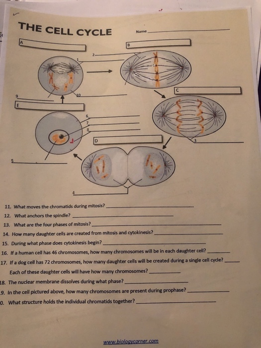
Solved THE CELL CYCLE 11. What moves the chromatids during
Nucleus
