Unraveling The Secrets Of Cell Division: Microtubules, The Master Movers
Microtubules are organelles that play a crucial role in cell division, particularly in moving chromatids around during the process. Chromatids are the individual strands of chromosomes, and they need to be accurately separated and distributed to the daughter cells during cell division to ensure genetic stability.
Microtubules are long, hollow cylinders made of tubulin protein subunits. They are highly dynamic structures that can quickly assemble and disassemble, allowing them to perform various functions in the cell. During cell division, microtubules form a spindle apparatus that extends from one pole of the cell to the other. The spindle fibers attach to the kinetochores of the chromosomes, which are specialized protein complexes located at the centromeres of the chromosomes. The kinetochores serve as the attachment points for the spindle fibers.
Once the spindle apparatus is fully assembled, the spindle fibers begin to shorten, pulling the chromosomes towards the opposite poles of the cell. This process is known as anaphase and is a critical step in ensuring that each daughter cell receives a complete set of chromosomes. Without microtubules and their ability to move chromatids, cell division would not be possible, and genetic information would not be accurately from one generation to the next.
"what moves chromatids around during cell division what organelle anchors
Microtubules are organelles that play a crucial role in cell division, particularly in moving chromatids around during the process. Chromatids are the individual strands of chromosomes, and they need to be accurately separated and distributed to the daughter cells during cell division to ensure genetic stability.
- Structure: Microtubules are long, hollow cylinders made of tubulin protein subunits.
- Function: Microtubules form a spindle apparatus that extends from one pole of the cell to the other and attaches to the kinetochores of the chromosomes.
- Dynamics: Microtubules are highly dynamic structures that can quickly assemble and disassemble, allowing them to perform various functions in the cell.
- Kinetochores: Kinetochores are specialized protein complexes located at the centromeres of the chromosomes and serve as the attachment points for the spindle fibers.
- Anaphase: Once the spindle apparatus is fully assembled, the spindle fibers begin to shorten, pulling the chromosomes towards the opposite poles of the cell.
- Chromatid Separation: Microtubules are responsible for accurately separating the chromatids, ensuring that each daughter cell receives a complete set of chromosomes.
- Cell Division: Without microtubules and their ability to move chromatids, cell division would not be possible, and genetic information would not be accurately passed from one generation to the next.
- Genetic Stability: Microtubules play a critical role in maintaining genetic stability by ensuring the accurate segregation of chromosomes during cell division.
In conclusion, microtubules are essential organelles that play a crucial role in cell division, particularly in moving chromatids around during the process. Their dynamic structure and ability to attach to kinetochores allow them to accurately separate and distribute chromosomes to daughter cells, ensuring genetic stability and the proper functioning of organisms.
Structure
The structure of microtubules is directly related to their function in moving chromatids around during cell division. Microtubules are long, hollow cylinders composed of tubulin protein subunits. This structure allows them to be highly dynamic and flexible, which is essential for their role in cell division.
- Polymerization and Depolymerization: Microtubules can quickly assemble and disassemble by adding or removing tubulin subunits from their ends. This dynamic behavior allows them to rapidly form and re-form the spindle apparatus during cell division.
- Polarity: Microtubules have a defined polarity, with a plus end and a minus end. The plus end is the site of tubulin addition and growth, while the minus end is the site of tubulin removal and disassembly. This polarity is crucial for the proper attachment of microtubules to the kinetochores of chromosomes and for the subsequent movement of chromatids.
- Rigidity: Microtubules are relatively rigid structures, which allows them to withstand the forces generated during chromosome segregation. This rigidity ensures that chromatids are accurately separated and distributed to the daughter cells.
- Motor Proteins: Microtubules interact with motor proteins, such as dynein and kinesin, which use the energy from ATP to move along microtubule tracks. These motor proteins play a crucial role in the movement of chromosomes during cell division.
In conclusion, the structure of microtubules, with their dynamic behavior, polarity, rigidity, and ability to interact with motor proteins, is essential for their function in moving chromatids around during cell division. This process is critical for ensuring the accurate segregation of chromosomes and the maintenance of genetic stability.
Function
The formation of the spindle apparatus is a crucial aspect of microtubule function in moving chromatids around during cell division. The spindle apparatus is a bipolar structure that extends from one pole of the cell to the other and is composed of microtubule fibers. The microtubule fibers interact with the kinetochores of chromosomes, which are specialized protein complexes located at the centromeres of the chromosomes.
The attachment of microtubule fibers to the kinetochores is essential for the accurate segregation of chromosomes during cell division. Once the spindle apparatus is fully assembled, the microtubule fibers begin to shorten, pulling the chromosomes towards the opposite poles of the cell. This process, known as anaphase, ensures that each daughter cell receives a complete set of chromosomes.
Without the formation of the spindle apparatus and the subsequent attachment of microtubule fibers to the kinetochores, the accurate segregation of chromosomes would not be possible. This would lead to genetic instability and could result in developmental abnormalities or diseases.
In conclusion, the function of microtubules in forming a spindle apparatus that extends from one pole of the cell to the other and attaches to the kinetochores of the chromosomes is fundamental to their role in moving chromatids around during cell division. This process ensures the accurate segregation of chromosomes and the maintenance of genetic stability.
Dynamics
The dynamic nature of microtubules is critical to their role in moving chromatids around during cell division. Microtubules can quickly assemble and disassemble, allowing them to rapidly form and re-form the spindle apparatus. This dynamic behavior is essential for the accurate segregation of chromosomes during cell division.
- Polymerization and Depolymerization: Microtubules can quickly assemble and disassemble by adding or removing tubulin subunits from their ends. This dynamic behavior allows them to rapidly form the spindle apparatus during cell division and then disassemble once the chromosomes have been segregated.
- Polarity: Microtubules have a defined polarity, with a plus end and a minus end. The plus end is the site of tubulin addition and growth, while the minus end is the site of tubulin removal and disassembly. This polarity is crucial for the proper attachment of microtubules to the kinetochores of chromosomes and for the subsequent movement of chromatids.
- Motor Proteins: Microtubules interact with motor proteins, such as dynein and kinesin, which use the energy from ATP to move along microtubule tracks. These motor proteins play a crucial role in the movement of chromosomes during cell division.
In conclusion, the dynamic nature of microtubules, including their ability to polymerize and depolymerize, their polarity, and their ability to interact with motor proteins, is essential for their role in moving chromatids around during cell division. This dynamic behavior ensures the accurate segregation of chromosomes and the maintenance of genetic stability.
Kinetochores
Kinetochores are essential for the proper segregation of chromosomes during cell division. They serve as the attachment points for the spindle fibers, which are responsible for pulling the chromosomes to opposite poles of the cell. Without kinetochores, the spindle fibers would not be able to attach to the chromosomes, and the chromosomes would not be able to segregate properly. This would lead to aneuploidy, a condition in which cells have an abnormal number of chromosomes. Aneuploidy can cause a variety of developmental problems, including birth defects, intellectual disability, and cancer.
Kinetochores are complex structures that are composed of a number of different proteins. These proteins work together to ensure that the kinetochore is properly attached to the spindle fibers and that the chromosomes are segregated correctly. The assembly and function of kinetochores is tightly regulated by the cell cycle. This ensures that kinetochores are only present at the centromeres of chromosomes during cell division.
The study of kinetochores is important for understanding the process of cell division. By understanding how kinetochores work, scientists can gain insights into how cells divide and how errors in cell division can lead to disease.
In conclusion, kinetochores are essential for the proper segregation of chromosomes during cell division. They serve as the attachment points for the spindle fibers, which are responsible for pulling the chromosomes to opposite poles of the cell. Without kinetochores, the spindle fibers would not be able to attach to the chromosomes, and the chromosomes would not be able to segregate properly. This would lead to aneuploidy, a condition in which cells have an abnormal number of chromosomes. Aneuploidy can cause a variety of developmental problems, including birth defects, intellectual disability, and cancer.
Anaphase
Anaphase is a critical stage of cell division during which the chromosomes are separated and pulled to opposite poles of the cell. This process is mediated by the spindle apparatus, a complex structure composed of microtubules. The shortening of the spindle fibers during anaphase generates the force necessary to separate the chromosomes.
- Kinetochore Attachment: The spindle fibers attach to the chromosomes at specialized protein complexes called kinetochores. These attachments ensure that the chromosomes are properly aligned and pulled apart during anaphase.
- Motor Proteins: The movement of chromosomes during anaphase is facilitated by motor proteins, such as dynein and kinesin. These proteins use the energy from ATP to move along microtubule tracks, pulling the chromosomes towards the opposite poles of the cell.
- Microtubule Dynamics: The shortening of the spindle fibers during anaphase is driven by the dynamic behavior of microtubules. Microtubules can rapidly assemble and disassemble, allowing them to shorten and pull the chromosomes apart.
- Chromosome Segregation: The ultimate goal of anaphase is to segregate the chromosomes into two separate sets, one for each daughter cell. This process ensures that each daughter cell receives a complete set of genetic information.
In conclusion, anaphase is a crucial stage of cell division during which the spindle apparatus shortens, pulling the chromosomes apart and ensuring the proper segregation of genetic material into two daughter cells.
Chromatid Separation
Chromatid separation is a crucial aspect of cell division, as it ensures that each daughter cell receives a complete set of genetic information. This process is carried out by microtubules, which are dynamic structures that form the spindle apparatus during cell division. The spindle apparatus consists of microtubule fibers that attach to the kinetochores of chromosomes, specialized protein complexes located at the centromeres of the chromosomes. Once the spindle apparatus is fully assembled, the microtubule fibers begin to shorten, pulling the chromosomes towards opposite poles of the cell. This process, known as anaphase, ensures that each daughter cell receives a complete set of chromosomes.
Understanding the connection between chromatid separation and microtubules is essential for comprehending the fundamental mechanisms of cell division. Without accurate chromatid separation, genetic information would not be properly distributed to daughter cells, which could lead to developmental abnormalities and genetic diseases. Therefore, the role of microtubules in chromatid separation is critical for maintaining genetic stability and ensuring the proper functioning of organisms.
In conclusion, chromatid separation is a fundamental process in cell division, carried out by microtubules and the spindle apparatus. This process ensures the accurate distribution of genetic information to daughter cells, maintaining genetic stability and the proper development and functioning of organisms.
Cell Division
Understanding the connection between cell division and microtubules is crucial in exploring the mechanisms that ensure the accurate segregation of genetic material during cell division. Microtubules, as the primary structures responsible for moving chromatids, play a pivotal role in the process. Without their ability to move chromatids, cell division would be disrupted, leading to genetic instability and potential developmental abnormalities.
- Role of Microtubules in Cell Division: Microtubules form the mitotic spindle, a dynamic structure that orchestrates the movement of chromosomes during cell division. They attach to the kinetochores of chromosomes, ensuring the proper alignment and segregation of genetic material into daughter cells.
- Consequences of Microtubule Dysfunction: Disruptions in microtubule function can lead to a variety of genetic abnormalities. Aneuploidy, a condition characterized by an abnormal number of chromosomes, can arise due to improper chromosome segregation caused by microtubule dysfunction. This can have severe consequences for cell viability and overall organismal health.
- Microtubules in Cancer: Microtubule dysfunction has been implicated in the development and progression of cancer. Many chemotherapeutic drugs target microtubules, disrupting their function and leading to cell death in rapidly dividing cancer cells.
- Microtubules in Meiosis: In the context of meiosis, the process of gamete formation, microtubules play a crucial role in ensuring the proper segregation of homologous chromosomes. Accurate chromosome segregation during meiosis is essential for maintaining genetic diversity and preventing genetic disorders in offspring.
In conclusion, the connection between cell division, microtubules, and the movement of chromatids is fundamental to our understanding of genetic stability and the proper functioning of cells. Microtubules, as the key players in this process, ensure the accurate segregation of genetic material during cell division, contributing to the maintenance of genetic integrity and the overall health of organisms.
Genetic Stability
The accurate segregation of chromosomes during cell division is essential for maintaining genetic stability. Genetic stability refers to the preservation of the correct number and structure of chromosomes throughout cell divisions. Microtubules, as the primary structures responsible for moving chromatids during cell division, play a critical role in ensuring genetic stability.
During cell division, microtubules form the mitotic spindle, a dynamic structure that orchestrates the movement of chromosomes. The mitotic spindle attaches to the kinetochores of chromosomes, specialized protein complexes located at the centromeres of the chromosomes. Once the mitotic spindle is fully assembled, the microtubule fibers begin to shorten, pulling the chromosomes towards opposite poles of the cell. This process, known as anaphase, ensures that each daughter cell receives a complete set of chromosomes.
If microtubules are disrupted or dysfunctional, the accurate segregation of chromosomes can be compromised, leading to genetic instability. Aneuploidy, a condition characterized by an abnormal number of chromosomes, can arise due to improper chromosome segregation caused by microtubule dysfunction. Aneuploidy can have severe consequences for cell viability and overall organismal health. For example, aneuploidy has been linked to developmental disorders, intellectual disabilities, and cancer.
In conclusion, microtubules play a critical role in maintaining genetic stability by ensuring the accurate segregation of chromosomes during cell division. Understanding the connection between microtubules and genetic stability is crucial for comprehending the fundamental mechanisms of cell division and the importance of maintaining genetic integrity for proper cell function and organismal health.
FAQs on "what moves chromatids around during cell division what organelle anchors"
This section addresses frequently asked questions related to the role of microtubules in moving chromatids during cell division.
Question 1: What is the primary structure responsible for moving chromatids during cell division?
Answer: Microtubules are the primary structures responsible for moving chromatids during cell division. They form the mitotic spindle, a dynamic structure that orchestrates the movement of chromosomes during cell division.
Question 2: How do microtubules attach to chromosomes?
Answer: Microtubules attach to chromosomes at specialized protein complexes called kinetochores, located at the centromeres of the chromosomes.
Question 3: What is the significance of accurate chromosome segregation during cell division?
Answer: Accurate chromosome segregation is essential for maintaining genetic stability. Disruptions in microtubule function can lead to aneuploidy, a condition characterized by an abnormal number of chromosomes, which can have severe consequences for cell viability and organismal health.
Question 4: What is the role of microtubules in maintaining genetic stability?
Answer: Microtubules play a critical role in maintaining genetic stability by ensuring the accurate segregation of chromosomes during cell division. They form the mitotic spindle, which facilitates the proper alignment and separation of chromosomes, ensuring that each daughter cell receives a complete set of genetic information.
Question 5: How does microtubule dysfunction affect cell division and genetic stability?
Answer: Microtubule dysfunction can disrupt cell division and compromise genetic stability. Impaired microtubule function can lead to improper chromosome segregation, resulting in aneuploidy and potential developmental abnormalities, intellectual disabilities, and cancer.
Question 6: What is the significance of microtubules in the context of cancer treatment?
Answer: Microtubules are important targets for cancer treatment. Many chemotherapeutic drugs target microtubules, disrupting their function and leading to cell death in rapidly dividing cancer cells.
Summary: Microtubules play a crucial role in moving chromatids during cell division, ensuring the accurate segregation of chromosomes and maintaining genetic stability. Understanding microtubule function is essential for comprehending the mechanisms of cell division and the importance of genetic stability for proper cell function and organismal health.
Transition to the next article section: This concludes our exploration of the role of microtubules in moving chromatids during cell division. In the next section, we will delve into the mechanisms that regulate microtubule dynamics and ensure the fidelity of chromosome segregation.
Tips for Understanding Microtubule Function in Cell Division
To enhance your understanding of microtubule function in cell division, consider the following tips:
Tip 1: Grasp the Dynamic Nature of MicrotubulesMicrotubules are not static structures but rather highly dynamic entities constantly assembling and disassembling. This dynamic behavior allows them to rapidly form and re-form the mitotic spindle during cell division.Tip 2: Comprehend Kinetochore FunctionKinetochores, specialized protein complexes, serve as the attachment points between microtubules and chromosomes. A proper understanding of kinetochore structure and function is crucial for grasping chromosome segregation mechanisms.Tip 3: Visualize Anaphase DynamicsAnaphase is a critical stage where microtubule shortening drives chromosome separation. Visualizing the spindle apparatus and the shortening microtubule fibers will aid in comprehending the mechanics of chromosome segregation.Tip 4: Explore Microtubule-Motor Protein InteractionsMotor proteins, such as dynein and kinesin, interact with microtubules and utilize cellular energy to facilitate chromosome movement. Understanding these interactions will deepen your knowledge of chromosome segregation.Tip 5: Examine Microtubule RegulationCell division is a tightly regulated process, and microtubule dynamics are no exception. Exploring the regulatory mechanisms that control microtubule assembly, disassembly, and attachment to chromosomes will provide a comprehensive understanding of cell division.Tip 6: Investigate Microtubule Dysfunction ConsequencesMicrotubule dysfunction can lead to chromosome segregation errors and aneuploidy. Understanding the consequences of microtubule dysfunction will reinforce the importance of microtubule function in maintaining genetic stability.Tip 7: Connect Microtubules to Cell Division and DiseaseMicrotubules are not only essential for cell division but also play a role in various cellular processes. Exploring the connections between microtubules, cell division, and diseases like cancer will broaden your perspective on microtubule function.Summary: By incorporating these tips into your learning, you will gain a deeper understanding of microtubule function in cell division and its implications for genetic stability and cellular health.Transition to the article's conclusion: This exploration of microtubule function in cell division highlights their critical role in ensuring accurate chromosome segregation and maintaining genetic stability. Understanding these concepts is fundamental for comprehending the mechanisms of cell division and their implications for cellular health and disease.
Conclusion
This comprehensive exploration of "what moves chromatids around during cell division what organelle anchors" has shed light on the crucial role of microtubules in cell division and genetic stability. Microtubules, as the primary structures responsible for moving chromatids, form the mitotic spindle apparatus, ensuring the accurate segregation of chromosomes during cell division.
The dynamic nature of microtubules, their interaction with kinetochores, and the regulation of their dynamics are essential for the fidelity of chromosome segregation. Disruptions in microtubule function can lead to aneuploidy, a condition with severe consequences for cell viability and organismal health. Understanding microtubule function is not only fundamental for cell biology but also has implications for understanding diseases like cancer.
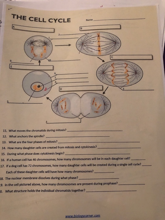
Solved THE CELL CYCLE 11. What moves the chromatids during
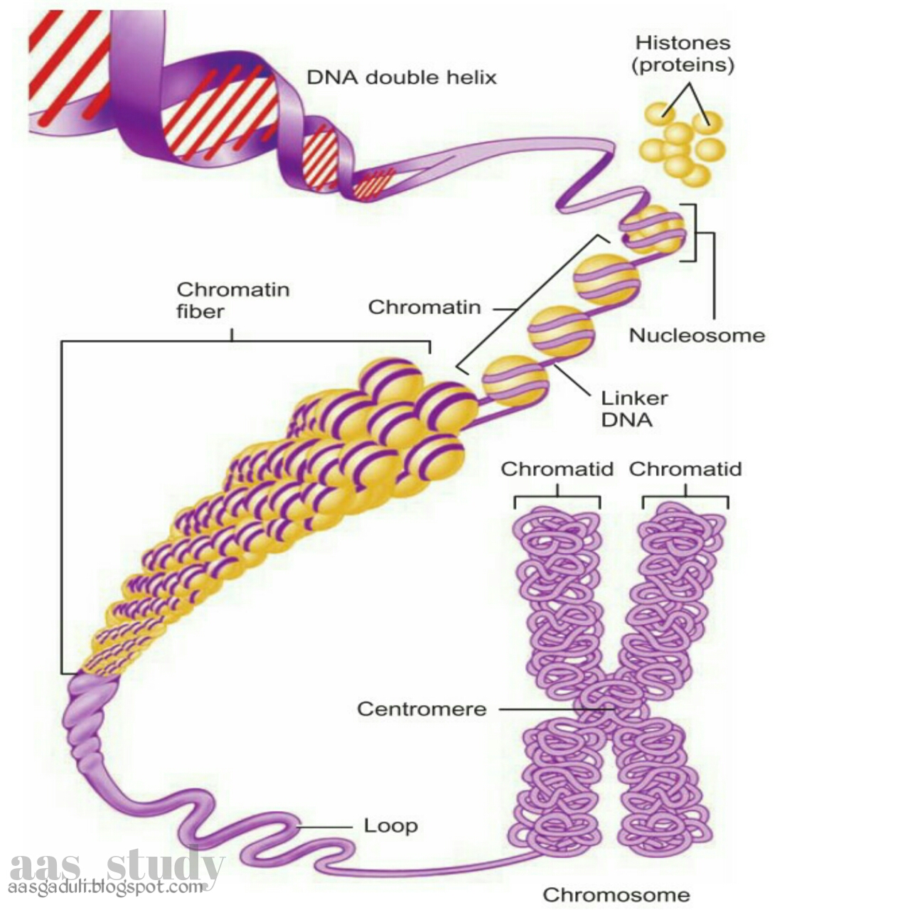
Nucleus
